Wednesday, February 28, 2007

Regulation of Alzheimer's disease amyloid-beta formation
.
Alzheimer's Donation
Donate Online Now
.


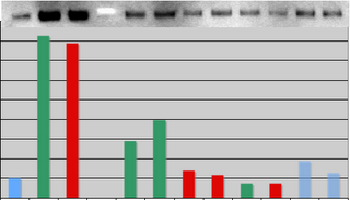
lzheimer's disease (AD) is associated with accumulation of the neurotoxic peptide amyloid-beta (A), which is produced by sequential cleavage of amyloid precursor protein (APP) by the aspartyl protease beta-secretase and the presenilin-dependent protease gamma-secretase. An increase of casein kinase 1 (CK1) expression has been described in the human AD brain. We show, by using in silico analysis, that APP, beta-secretase, and gamma-secretase subunits contain, in their intracellular regions, multiple CK1 consensus phosphorylation sites, many of which are conserved among human, rat, and mouse species. Overexpression of constitutively active CK1, one of the CK1 isoforms expressed in brain, leads to an increase in A peptide production. Conversely, three structurally dissimilar CK1-specific inhibitors significantly reduced endogenous A peptide production. By using mammalian cells expressing the C-terminal fragment of APP, it was possible to demonstrate that CK1 inhibitors act at the level of gamma-secretase cleavage. Importantly, Notch cleavage was not affected. Our results indicate that CK1 represents a therapeutic target for prevention of A formation in AD.

The studies lead author Marc Flajolet and from the Nobel Prize winning laboratory of Paul Greengard, director of the Fisher Center for Alzheimer's Disease Research at Rockefeller. "Studies of brain tissue from Alzheimer's patients have shown an increase in casein kinase 1 expression," says Greengard, Vincent Astor Professor and head of the Laboratory of Molecular and Cellular Neuroscience. "We found that the key enzymes involved in beta-amyloid production - called BACE and gamma-secretase - were targets of casein kinase 1, so we investigated what role it might be playing." The scientists modified mouse cells to generate a form of casein kinase 1 that was always active, and found that these cells produced more beta-amyloid protein than normal. Then, using three different types of chemicals, they blocked the protein from functioning. When they did this, they were able to reverse the production of beta-amyloid protein, without affecting the signaling of Notch proteins. The studies suggest that an Alzheimer's therapy based on these chemicals could reduce or halt beta-amyloid build-up without causing side effects. "Numerous efforts have been directed at the development of drugs that inhibit gamma-secretase," says Greengard, "but there have been significant side effects in animal studies. Our hope is that this research might lead to drugs that don't have those problems."

Tuesday, February 27, 2007

Grandma may be better advised
.
Alzheimer's Donation
Donate Online Now
.


Earlier this year, Nintendo launched the American version of a game for seniors called "Brain Age: Train Your Brain in Minutes a Day." Developed in collaboration with Japanese neurologist Ryuta Kawashima, this game is based on the idea that regular mental exercises—such as Sudoku puzzles, Stroop tasks and simple math problems—can keep aging brains sharper. Similar games are being developed by other companies intent on capturing this new market. Radica, a subsidiary of Mattel, is releasing "Brain Games," word and memory games based on research at the University of California at Los Angeles Center of Aging. Another company, MindFit, is also marketing cognitive training games for seniors, citing extensive evidence for the 'use it or lose it' theory of cognitive decline with aging. Despite the marketing hype, there is no conclusive evidence that mental games alone slow brain aging.

As people age, their performance declines on many tasks, particularly those involving working memory, and they lose some brain volume. Older people also show less activation than younger adults in brain imaging studies during a wide variety of tasks. Positron emission tomography shows broader recruitment of different brain areas in older adults during specific tasks. However, it is unclear whether this change reflects failure to recruit specific neural processes or compensation for lost neural function.

Animal studies indicate that environmental factors such as enrichment or physical exercise may affect cognitive function and slow down certain markers of aging. Extrapolating the results to humans, though challenging, seems to confirm this trend for physical fitness. A meta-analysis of aerobic fitness training studies in aging found significant effects on cognition, particularly on tasks involving executive control processes. Recently, a six-month randomized clinical trial split older adults into two groups, one that received aerobic training and another that did stretching and toning (nonaerobic) exercises. After training, the aerobic group showed increases in both gray and white matter volumes. However, the consequences of increased volume for cognition are not conclusive, and there remains no clear evidence correlating different levels of physical activity to performance.

The data linking mental training to amelioration of age-related cognitive decline are murkier. Most people, young or old, improve their performance on tasks with practice. Some studies suggest that cognitive training improves mental ability. A randomized control trial with 2,832 seniors reported that as few as ten sessions of cognitive training improved performance on tested tasks even five years later. Participants also reported a higher confidence in being able to handle their day-to-day tasks, though self-report is not the most reliable measure of improvement in real-life activities or ability to live independently. In another randomized controlled study, adults in the Posit Science Training program, who performed increasingly difficult auditory stimulus recognition, discrimination and memory tasks, showed improvements in the specific tasks as well as on a global auditory memory test. However, other studies have found little evidence for a slower rate of age-related decline in reasoning in individuals who spend more time doing mentally stimulating tasks such as crossword puzzles.

Most scientists would argue that there is no harm in keeping physically fit or cognitively challenged, and that the only downside to such programs is a dent in your wallet. Still, there is an inherent danger in conferring scientific credibility on products that may not live up to their claims. Instead of investing in these cognitive programs (or even a future video game akin to the Nintendo Wii that combines physical activity with mental exercise), grandma may be better advised to take regular walks, stay socially active and challenge her mind without the aid of commercial games.
Nature Neuroscience - 10, 263 (2007)
doi:10.1038/nn0307-263

Monday, February 26, 2007

The bill seeks to find cure for Alzheimer's disease
.
Alzheimer's Donation
Donate Online Now
.


The Alzheimer's Association strongly backs H.R. 1032, the Alzheimer's Treatment and Caregiver Support Act, introduced Feb. 13 by Rep. Maxine Waters (D-Calif.). The bill seeks to find prevention methods, treatments and an eventual cure for Alzheimer's disease while also providing much needed support for those with the disease and their caregivers. "The Alzheimer's Treatment and Caregiver Support Act offered by Rep. Maxine Waters is a tremendous step in creating awareness about Alzheimer's disease, which is currently affecting 4.5 million Americans over the age of 65," said Harry Johns, Alzheimer's Association president and CEO. "With that number expected to soar to as many as 16 million by 2050, this legislation addresses the need for education, training, support and tangible services to assist those with the disease and their families.

"The Alzheimer's Association applauds Rep. Maxine Waters for continuing to make Alzheimer's a priority and for championing the expansion of community-based services, especially in underserved communities, to assist those in need." Last year, Rep. Waters led the fight to restore funding for the Alzheimer's Association Safe Return® program that helps reunite loved ones with a person with dementia who has wandered. The Alzheimer's Association is appreciative of Rep. Waters' leadership on this issue and will be working with advocates to ensure passage of H.R. 1032 in the House of Representatives.

Sunday, February 25, 2007

Cholesterol In Alzheimer's Disease
.
Alzheimer's Donation
Donate Online Now
.


The lower your cholesterol, the better your heart health. But a man's heart and his head don't always agree. In fact, the relationships among cholesterol levels, psychological function, and neurologic disorders are complex and sometimes controversial, reports the March 2007 issue of Harvard Men's Health Watch.

There are two major forms of dementia: vascular dementia and Alzheimer's disease. Vascular dementia results when blood vessel damage deprives the brain of oxygen. Brain cells die as a result, and mental function suffers. Some studies link high cholesterol levels to an increased risk of cognitive impairment, but others report the opposite. More research is needed to sort this out, but even now, investigations of HDL (good) cholesterol and mental function have consistently reported that high HDL levels appear to help preserve mental function in older people.

The connection between Alzheimer's disease and cholesterol is even more complex. Scientists have learned much of the damage of Alzheimer's comes from deposits of a sticky protein, called beta-amyloid, in vital areas of the brain. In some studies, high cholesterol levels appear to accelerate the formation of beta-amyloid plaques. People with the genetic trait that increases the level of a particular cholesterol transport protein have a greatly increased risk of late-onset Alzheimer's.

The urgent question is whether cholesterol-lowering drugs, such as statins, can reduce the risk of Alzheimer's disease. In the most recent studies, people who took statins did not appear to be at lower risk for the disease. Additional research is under way. Right now, it is too early for firm conclusions on the relationships among cholesterol, cognitive function, and statin therapy.

Saturday, February 24, 2007

Cut Alzheimer's Risk
.
Alzheimer's Donation
Donate Online Now
.


The benefits of Antioxidant Loaded Juice
Studies have shown that people who drink three or more servings of antioxidant loaded juice weekly are less prone to develop Alzheimer's disease! According to research found by a study in The American Journal of Medicine people who consumed more than one glass of juice per week were 76% less likely to develop Alzheimer's disease.

The Benefits of Taking Fish Oil
The benefits of supplementing your diet with omega 3 fatty acids are overwhelmingly positive. And, supplementing with fish oil makes even more sense since a lack of these omega 3 fatty acids has been linked to pre-menstrual syndrome, depression, bipolar disorder, schizophrenia, violence and suicide. But a recent survey showed that 85% of Americans are deficient in these fatty acids. And, 98% of pregnant and lactating women get less than 18% of the NIH's recommended amount. That's appalling!
How about this one? Nearly 20% of the American population is so low in DHA and EPA (two of the omega 3 fatty acids) that test methods are not able to detect any in their blood. Get started eating more fatty fish (like salmon, tuna, herring, sardines and anchovies) and start taking a high quality, pure fish oil supplements on a daily basis.

Alzheimer’s Disease and Parkinson’s Disease: a 360° view Nature Publishing Group has gathered together a collection of top quality research papers on Alzheimer’s Disease and Parkinson’s Disease from an assortment of our highly rated journals for you to preview at no charge. From genetics and behaviour to pharmacology, pharmcogenomics and brain circulation, NPG continues to publish top-tier research spanning the breadth of Alzheimer’s Disease and Parkinson’s Disease. Visit www.nature.com/360.

Friday, February 23, 2007

Cut the risk of developing Alzheimer's
.
Alzheimer's Donation
Donate Online Now
.
Older adults whose total folate intake is at or above the recommended dietary allowance (RDA) could cut their risk of developing Alzheimer's disease by more than half. The study, reported in the inaugural issue of the journal Alzheimer's & Dementia: The Journal of the Alzheimer’s Association, is the largest to date to show that the B vitamin folate could prevent Alzheimer's. According to the researchers, study participants who reported intake at or above the 400 microgram recommended daily allowance of folate had a 55 percent reduction in risk in developing Alzheimer’s. Investigators also noted that most people in the study who reached the recommended intake level did so by taking folate supplements, suggesting many people do not get the recommended amount of folate in their diet.


Folate is a B-vitamin nutrient found in foods such as leafy green vegetables, asparagus, broccoli, liver, oranges, and many types of beans and peas. Only 13% of study participants reached the recommended dietary allowance of folate from food alone. Epidemiologists at the University of California collected dietary information from 579 people age 60 and older who showed no sign of Alzheimer's. The volunteers kept track of the food they consumed during a typical week and told the researchers if they were taking vitamin supplements that contained folate. The researchers calculated the total amount of folate consumed and kept track of the recruits for an average of more than 9 years. During that time, 57 people developed Alzheimer's.


Thursday, February 22, 2007

Dementia drug development guidelines
.
Alzheimer's Donation
Donate Online Now
.

Live telecast
Product Description: On March 9, 2000, FDA's Psychopharmacologic Drugs Advisory Committee will discuss drug development for Alzheimer's and other dementias. The panel will address the problem of how to identify, define, and name clinical entities that fall under the dementia category in order to prevent misleading labeling. The committee's deliberations will be televised live by videoconferencing, webcast and telephone link ups. In addition, videotapes will be available shortly after the meeting.
For information on how to receive the live telecast, use the Request Free Product Information online form above or contact: Request Free Product Information

Wednesday, February 21, 2007

Alzheimer's disease is a synaptic disease
.
Alzheimer's Donation
Donate Online Now
.


Alzheimer's disease (AD) was first described in 1907 by Alois Alzheimer in a 51-year-old woman with the following symptoms: progressive memory impairment; disordered cognitive function; altered behavior, including paranoia and delusion; and decline in language function. The brains of AD subjects are characterized by accumulation of neuritic plaques containing amyloid protein and neurofibrillary tangles, accompanied by loss of certain types of receptors and neurons. Pathological changes begin in the transentorhinal allocortex with degeneration accentuated in the hippocampal formation and entorhinal, frontal, and temporal cortices.
It is known that Alzheimer's disease (AD) is a synaptic disease that involves various neurotransmitter systems, particularly those where synaptic transmission is mediated by acetylcholine or glutamate (Glu). Nevertheless, very little is known about the properties of neurotransmitter receptors of the AD human brain. We have shown previously that cell membranes, carrying neurotransmitter receptors from the human postmortem brain, can be transplanted to frog oocytes, and their receptors will still be functional. Taking advantage of this fact, we have now studied the properties of Glu receptors (GluRs) from the cerebral cortices of AD and non-AD brains and found that oocytes injected with AD membranes acquired GluRs that have essentially the same functional properties as those of oocytes injected with membranes from non-AD brains. However, the amplitudes of the currents elicited by Glu were always smaller in the oocytes injected with membranes from AD brains. Western blot analyses of the same membrane preparations used for the electrophysiological studies showed that AD membranes contained significantly fewer GluR2/3 subunit proteins. Furthermore, the corresponding mRNAs were also diminished in the AD brain. Therefore, the smaller amplitude of membrane currents elicited by Glu in oocytes injected with membranes from an AD brain is a consequence of a reduced number of GluRs in cell membranes transplanted from the AD brain. Thus, using the comparatively simple method of microtransplantation of receptors, it is now possible to determine the properties of neurotransmitter receptors of normal and diseased human brains. That knowledge may help to decipher the etiology of the diseases and also to develop new treatments.

Tuesday, February 20, 2007

The largest research projects in the United States
.
Alzheimer's Donation
Donate Online Now
.
Almost all common diseases and health conditions are linked both to genetic and environmental factors, according to the researchers. "Understanding the critical interaction between genes and the environment on health will have an important impact on the way all of us look at health and disease in the future," said Schaefer. "For instance, our research could help us identify not only what diseases a person may be at risk for, but also identify how to reduce that risk, or how best to treat the disease. This research program provides an exciting opportunity to make significant progress toward improvements in health and medical care."
The Kaiser Permanente Northern California Division of Research is launching one of the largest research projects in the United States to examine the genetic and environmental factors that influence common diseases such as heart disease, cancer, diabetes, high blood pressure, Alzheimer's disease, asthma and many others. The goal of the Kaiser Permanente Research Program on Genes, Environment and Health (RPGEH) is to discover which genes and environmental factors -- the air we breathe, the water we drink, as well as lifestyles and habits -- are linked to specific diseases.
Participation in the RPGEH is completely voluntary. An individual's genetic information will not be used in genetic studies without their written consent. Moreover, as with all studies carried out by the Division of Research, protecting the confidentiality and security of our members' information is our first priority. Kaiser Permanente has a long track record of protecting the security and confidentiality of information provided by research participants. The KP Division of Research maintains separate information and databases from the health plan and members' medical records. No research data is stored with identifying information. In the database, each participant will be given a unique code that will replace their medical record number, name and other identifying information. The code can only be linked back to identifying information by a small number of Division of Research staff members who must have a special password. The data is for research only.

Monday, February 19, 2007

Structure and function in the brains of Alzheimer's
.
Alzheimer's Donation
Donate Online Now
.

A completely new approach to the study of Alzheimer's disease, initiated by a professor at the University of California, Santa Barbara, may solve a critical piece in the puzzle of the disease. This tragic neurological illness progressively erases memory in its millions of victims. The key to the new approach is understanding the way certain proteins in the brain fold, or rather "misfold."
Michael Bowers, a professor in the Department of Chemistry and Biochemistry, developed this project, which is being funded by the National Institutes of Health. Bowers is using specialized chemical research methods and applying them to biology. His research will depend upon the study of rare peptides, or strings of amino acids, that are difficult to produce. These will be provided by co-investigator David Teplow, a professor at UCLA's David Geffen School of Medicine, who has been involved in Alzheimer's research for over 10 years. Joan-Emma Shea, also a professor in UCSB's Department of Chemistry and Biochemistry, heads the theoretical modeling aspect of the project. He explained that now the hunt is on for the "small stuff." Because of their expertise in certain chemical methodologies, Bowers and his research group are able to track down the molecular level changes that lead to development of the disease.
The process of aggregation of proteins that cause the plaque begins in a way that Bowers has begun to clarify. The goal is to find non-toxic drugs that will interrupt the aggregation process. "If we can do that, we can stop the disease," said Bowers. "However, once you start losing neurons, things become very difficult, because the body doesn't readily replace them due to their very large size. If we could find a marker, early on, to indicate when the patient first has the disease, then the new drug or drugs that we hope to develop could prevent further damage." Bowers described his approach as a whole new way to determine the structure and composition of the Abeta 42 peptide and its oligomers that are primarily responsible for Alzheimer's disease. The research team is analyzing the way this peptide folds, causing it to aggregate and disrupt neuronal function.
The key aspect of ion mobility is its ability to measure accurate cross sections of complex aggregations of proteins and obtain information on their three-dimensional shape. When coupled with mass spectrometry, electrospray ionization, and high-level molecular modeling, it becomes a very powerful technique. The experiment starts with electrospray ionization, a method of spraying the solution containing the peptides of interest into fine droplets and then letting the droplets evaporate. Following evaporation, mass spectrometry is employed to determine the mass or weight of the species that were in the solution, and from that to determine the composition. Finally ion mobility is used to show the shape of the Abeta 42 peptide and its oligomers.

Sunday, February 18, 2007

Brain can manufacture fresh brain cells
.
Alzheimer's Donation
Donate Online Now
.


New evidence shows that the human brain can made fresh brain cells, researchers said in a study that may lead to better ways to treat brain damage and disease. It is known that other animals, such as rats and mice, make new brain cells throughout their lives and there had been indirect evidence that humans being can, too. Using MRI, scans and electron microscope images of tissue donated from the brains of people who died, Maurice Curtis of the University of Auckland in New Zealand and Peter Eriksson of Sahlgrenska Academy in Goteborg, Sweden, and colleagues found the elusive cells. Just as in mice and rats, these cells are born in one part of the brain and then migrate to the olfactory bulb, where smells are processed. They mature into neurons on the way. In animals, they said, brain damage prompts the birth of new cells. "Our study provides the foundation for this possibility in the adult human brain," they wrote in their report, published in the journal Science. Humans have far more developed brains, so searching for these cells has been harder than it was in rodents.
"This opens another direction by which we may discover ways to repair human brains that are damaged from injury or diseases, and underscores the importance of animal research in guiding biomedical research in humans," Baxter said. Another expert, Sebastian Brandner, head of the Division of Neuropathology at the Institute of Neurology at University College London, agreed. "These findings are important for several reasons: Understanding stem cell biology is essential to study brain repair in neurodegenerative diseases such as Alzheimer's and it is even possible that stem cells are the source of some brain tumors," Brandner said in a statement.

Saturday, February 17, 2007

Inhibitor of Alzheimer's Tangles
.
Alzheimer's Donation
Donate Online Now
.


Friday, February 16, 2007

Researchers Testing Alzheimer's
.
Alzheimer's Donation
Donate Online Now
.


A U.S. scientist says the true cause of the neurological damage resulting from Alzheimer's disease might rest in the way certain brain proteins fold.Professor Michael Bowers of the University of California-Santa Barbara says the key to his approach is understanding the way those proteins fold, or rather, "misfold."Bowers says until about five or six years ago, everyone assumed large amyloid plaques found in the brains of Alzheimer's victims caused the disease. "However, recent scientific discoveries indicate that these large, insoluble aggregates might merely be markers of the disease -- they do not cause the disease," said Bowers. "Rather, smaller soluble oligomers, or peptide complexes, are now felt to be the causative agents, and I find that very interesting." His research team is analyzing the structure and composition of the Abeta 42 peptide and its oligomers, studying the way that peptide folds, causing it to aggregate and disrupt neuronal function.
It could be one of the biggest scientific breakthroughs ever, a new way to treat and even prevent Alzheimer's disease. For a 70-year-old, Ada English is in good shape. She has no outward signs she has early stage Alzheimer's disease, except to her family. Ada's husband of 50 years, Joseph says it's subtle, but significant. The confusion and memory loss that comes and goes. Alzheimer's is caused by the build up of plaque that slowly destroys parts of the brain and until now there's been no effective treatment. But now researchers are testing a promising new vaccine. Preliminary research shows the intravenous drug can eliminate plaque and prevent it from forming.

Thursday, February 15, 2007

Neurotransmitter receptors of normal and Alzheimer's brains
.
Alzheimer's Donation
Donate Online Now
.

It is known that Alzheimer's disease (AD) is a synaptic disease that involves various neurotransmitter systems, particularly those where synaptic transmission is mediated by acetylcholine or glutamate (Glu). Nevertheless, very little is known about the properties of neurotransmitter receptors of the AD human brain. We have shown previously that cell membranes, carrying neurotransmitter receptors from the human postmortem brain, can be transplanted to frog oocytes, and their receptors will still be functional. Taking advantage of this fact, we have now studied the properties of Glu receptors (GluRs) from the cerebral cortices of AD and non-AD brains and found that oocytes injected with AD membranes acquired GluRs that have essentially the same functional properties as those of oocytes injected with membranes from non-AD brains. However, the amplitudes of the currents elicited by Glu were always smaller in the oocytes injected with membranes from AD brains. Western blot analyses of the same membrane preparations used for the electrophysiological studies showed that AD membranes contained significantly fewer GluR2/3 subunit proteins. Furthermore, the corresponding mRNAs were also diminished in the AD brain. Therefore, the smaller amplitude of membrane currents elicited by Glu in oocytes injected with membranes from an AD brain is a consequence of a reduced number of GluRs in cell membranes transplanted from the AD brain.
Thus, using the comparatively simple method of microtransplantation of receptors, it is now possible to determine the properties of neurotransmitter receptors of normal and diseased human brains. That knowledge may help to decipher the etiology of the diseases and also to develop new treatments.

Wednesday, February 14, 2007

The whereabouts of Alzheimer's patients
.
Alzheimer's Donation
Donate Online Now
.

A pair of shoes embedded with global positioning system (GPS) technology can be used to detect the whereabouts of Alzheimer's patients and other wearers anywhere in the world.

The Quantum Satellite Technology (QST) shoes can be adapted to constantly detect the whereabouts of Alzheimer's and dementia patients, emergency medical service (EMS) personnel, children, or any other person whose location needs to be known constantly. In the event of any concern, distress, or danger, the wearer simply presses a button hidden near the shoe's lace to activate a covert alarm locator that sends a distress signal. Additionally, if a concern arises about the shoe wearer’s safety, their location can be identified by placing a phone call to a 24-hour monitoring service.
The shoe can also store information about the wearer’s movements and vital measurements such as heart rate, body temperature, and stress levels, useful in case of a medical emergency. An integrated Bluetooth wireless connection can be configured to make wireless telephone calls, enabling the wearer to call for help and give his precise location.
The electronics are embedded in a special module cavity containing the GPS transmitter, an antenna, and the wireless communication system. The shoe is equipped with a mini universal serial bus (USB) connection, which serves as both a power plug for the rechargeable battery and for data transfer. The internal data device can store up to 100,000 records, including a complete medical profile of the wearer, and all the tracking and monitoring information recorded.

The QST shoes are made by Isaac Daniel (New York, NY, USA) and are currently available in a limited edition of 1,000 pairs.

Tuesday, February 13, 2007

Hippocampal damage impairs memory
.
Alzheimer's Donation
Donate Online Now
.

The hippocampus and adjacent medial temporal lobe structures are known to support declarative memory, but there is not consensus about what memory functions the hippocampus might support that are distinct from the functions of the adjacent cortex. One idea is that the hippocampus is specifically important for allocentric spatial memory, e.g., the hippocampus is especially needed to remember object locations when there is a shift in viewpoint between study and test. We tested this proposal in two experiments. Patients with damage limited to the hippocampus were given memory tests for object locations in a virtual environment. In the first experiment, participants studied locations of a variable number of images (one to five) and tried to remember the image locations from either the same viewpoint as during study (shift of 0°) or a different viewpoint (shift of 55°, 85°, or 140°). In each viewpoint condition (shifts of 0°, 55°, 85°, and 140°), patients performed normally when remembering one or two image locations. Further, performance declined to a similar degree in each viewpoint condition as patients tried to remember increasing numbers of image locations. In the second experiment, participants tried to remember four images after viewpoint shifts of 0°, 55°, 85°, or 140°. Patients were mildly impaired at all conditions (shifts of 0°, 55°, 85°, and 140°), and the impairment was no greater when viewpoint shifted. We conclude that damage to the hippocampus does not selectively impair viewpoint-independent spatial memory. Rather, hippocampal damage impairs memory as the memory load increases.
Several studies have found a connection with dementia, a range of conditions including Alzheimer's that -- over many years -- destroys memory, organized thinking, and eventually everyday functioning. One particularly striking study found that 60 percent of hospitalized patients with delirium were diagnosed with dementia over the next three years, compared with 18 percent of those without, according to a review of the research by James C. Jackson , a neuropsychologist at Vanderbilt University in Nashville.

Monday, February 12, 2007

A positron emission tomography
.
Alzheimer's Donation
Donate Online Now
.

A microdosing study with an investigational, carbon 11–labeled antiamyloid drug, 1,1'-methylene-di-(2-naphthol) (ST1859), and positron emission tomography (PET) in healthy volunteers (n = 3) and patients with Alzheimer's disease (n = 6).
The study aimed to assess the distribution and local tissue pharmacokinetics of the study drug in its target organ, the human brain. Before PET studies were performed in humans, the toxicologic characteristics of ST1859 were investigated by an extended single-dose toxicity study according to guidelines of the Food and Drug Administration and European Medicines Agency, which are relevant for clinical trials with a single microdose. After intravenous bolus injection of 341 21 MBq [11C]ST1859 (containing <11.4>

Sunday, February 11, 2007

Vanderbilt University: juices cut the chances of Alzheimer
.
Alzheimer's Donation
Donate Online Now
.

One of the key characteristics of people with the condition is the formation of clumps, or 'plaques' of beta amyloid proteins which are thought to destroy brain cells.

Scientists increasingly believe diet and lifestyle may affect the build up of these damaging proteins.

Studies have found a Mediterranean-style diet rich in plant foods and fish and low in red meat cuts the risk of developing the brain disease by up to two-thirds.

Research in mice has also found that those given high-cholesterol diets have more amyloid beta proteins in their brain.

And there is growing evidence that taking cholesterol-lowering statins makes people less likely to develop Alzheimer's later in life.

To understand what lay behind this trend, Dr Brett Garner, of the Prince of Wales Medical Research Institute in Sydney, Australia, and his colleagues used human and animal cells to probe how brain cells regulate their levels of cholesterol.

In the arteries it is known that ABC proteins help control cholesterol levels by expelling it from the immune cells.

The study, reported in the Journal of Biological Chemistry found these proteins were also present in the brain cells.

When the boosted levels of the proteins by tweaking genes that affect production, cell lines production of amyloid beta protein fell.

The study also identified another protein in brain cells called apoE that regulates cholesterol removal from brain cells.

Dr Garner told New Scientist magazine that drugs that increase expression of these proteins might slow the progression of Alzheimer's.

Similar drugs are already being used for research into heart disease.

He said: "A lot of people think there could be converging factors involved in these diseases."

Large amounts of harmful cholesterol are found in foods high in saturated fats such as red meat, butter, cheese and offal such as liver and kidneys.

If people have a diet high in saturated fats, their liver produces more of the harmful form of cholesterol called LDL, which is linked to a higher risk of heart disease and stroke.

Scientists increasingly believe an unhealthy diet may be a contributing factor in developing dementia.

Previous research has found fish oil capsules may help slow the mental decline of those with very mild Alzheimer's disease.

Last September a team from Vanderbilt University in Tennessee found drinking fruit and vegetable juices more than three times a week could dramatically cut the chances of developing the condition.

Researchers from who followed almost 2,000 volunteers for up to ten years found the risk of Alzheimer's was 76 per cent lower for those who drank juices more than three times a week compared with those who drank them less than once a week.

Japanese scientists also found last year that green tea could halve the risk of mental decline in old age.

They found those who drank the tea the most - more than two cups a day - had a 54 per cent lower risk of dementia than those who drank the least.

Check out Publix GreenWise Market to find a variety of wholesome, natural and organic products for healthier living.

Kroger Cares about your health and well being! Click here to check out!

Saturday, February 10, 2007

Anesthetics may produce Alzheimer's-like changes in the brain
.
Alzheimer's Donation
Donate Online Now
.

Alzheimer's disease is characterized by plaques within the brain of amyloid-beta protein (A-beta), which is toxic to brain cells. A-beta is formed when the larger amyloid precursor protein (APP) is clipped by two enzymes -- beta-secretase, also known as BACE, and gamma-secretase -- to release the A-beta fragment. Normal processing of APP by an enzyme called alpha-secretase produces an alternative, non-toxic protein.

A new study has found how one of the most commonly used anesthetics may produce Alzheimer's-like changes in the brain. Previous studies have shown that applying the anesthetic isoflurane to cultured neural cells can lead to generation of amyloid-beta protein -- the key component of senile plaques seen in the brains of Alzheimer's patients -- and to the cell-death process known as apoptosis. In the Feb. 7 Journal of Neuroscience, researchers from Massachusetts General Hospital (MGH) and colleagues describe how isoflurane may set off a process in which A-beta generation and apoptosis interact with and magnify each other. Since this work was done in cell cultures, it is unknown whether the findings reflect a possible effect of the anesthetic on human brains.
Some studies have indicated that general anesthesia may increase the risk of developing Alzheimer's disease. It also is known that a small but significant number of surgical patients experience a form of dementia in the postoperative period, but there is insufficient evidence of a direct connection between anesthesia and the risk of dementia. Previous articles -- including a recent report from the same research team -- have shown that isoflurane increases both A-beta generation and apoptosis in several types of cultured cells. The current study was designed to investigate the relationship between isoflurane-induced apoptosis and A-beta generation.
In a series of experiments, the researchers first found that applying isoflurane to cultured neural cells increased the activation of the enzyme caspase -- a key player in a pathway leading to apoptosis -- with no change in A-beta generation or APP processing. When they applied isoflurane to neural cells that express APP and had been treated with a caspase inhibitor, the expected changes in APP processing and A-beta generation were significantly reduced, indicating that caspase activation is essential to the pathway leading to A-beta generation and aggregation.
The researchers also found that isoflurane appears to raise levels of the A-beta-releasing enzymes BACE and gamma secretase and that generation of A-beta plaques further increases isoflurane-induced caspase activation. In addition, adding A-beta to neuronal cells that do not express APP also increased caspase activation in response to isoflurane. Overall, the study's results define molecular pathways by which isoflurane induces deposition of A-beta, both directly and via caspase activation, and by which A-beta deposits lead to further caspase activation and apoptosis.

Friday, February 09, 2007

Inhibitors of Amyloid beta aggregation prevent and reverse Alzheimer
.
Alzheimer's Donation
Donate Online Now
.

When given orally to a transgenic mouse model of Alzheimer disease, cyclohexanehexol stereoisomers inhibit aggregation of amyloid peptide (A) into high-molecular-weight oligomers in the brain and ameliorate several Alzheimer disease–like phenotypes in these mice, including impaired cognition, altered synaptic physiology, cerebral A pathology and accelerated mortality. These therapeutic effects, which occur regardless of whether the compounds are given before or well after the onset of the Alzheimer disease–like phenotype, support the idea that the accumulation of A oligomers has a central role in the pathogenesis of Alzheimer disease.
Multiple lines of evidence suggest that the accumulation of neurotoxic oligomeric aggregates of A may be a central event in the pathogenesis of Alzheimer disease1, 2. If correct, this hypothesis predicts that inhibitors of A aggregation and toxicity may be effective in blocking this pathogenic cascade. We report here that orally administered cyclohexanehexol stereoisomers can block the accumulation of A oligomers in a dose-dependent manner, and reduce Alzheimer disease–like behavioral deficits, Alzheimer disease–like neuropathology and accelerated mortality in a transgenic mouse model of Alzheimer disease. These in vivo experimental results both strongly support the notion that accumulation of A oligomers has a significant role in the pathogenesis of Alzheimer disease and suggest that inhibition of A aggregation may be a useful therapy for this disease.

Thursday, February 08, 2007

Missing link in Alzheimer’s disease
.
Alzheimer's Donation
Donate Online Now
.

Scientists at the University of Virginia have identified what appears to be a major missing link in the process that destroys nerve cells in Alzheimer’s disease, an incurable disease that slowly destroys memory and cognitive abilities. The findings are reported in the Nov. 20, 2006, issue of the Journal of Cell Biology and could eventually lead to new drugs that target and disrupt specific proteins that conspire in the brain to cause Alzheimer’s.

In Alzheimer’s disease, two kinds of abnormal structures accumulate in the brain: amyloid plaques and neurofibrillary tangles. The plaques contain fibrils that are made from protein fragments called “beta-amyloid peptides.” The tangles also are fibrous, but they are made from a different substance, a protein called “tau.” In the new U.Va. study, the researchers found a deadly connection between beta-amyloid and tau, one that occurs before they form plaques and tangles, respectively.

According to George Bloom, the senior author of the study and a professor of biology and cell biology at U.Va., this connection causes the swiftest, most sensitive and most dramatic toxic effect of beta-amyloid found so far. What makes it most remarkable, though, is that it requires a form of amyloid that represents the building blocks of plaques, so called “pre-fibrillar beta-amyloid,” and it only happens in cells that contain tau. Even though they account for just ~10 percent of the cells in the brain, nerve cells are the major source of tau, which likely explains why they are specifically attacked in Alzheimer’s disease.

The researchers used cultured mammalian cells that either did or did not make tau to study how cells respond to beta-amyloid. They found that pre-fibrillar, but not fibrillar beta-amyloid works together with tau to break apart microtubules — highways along which “synapse” replacement parts move rapidly in the nerve cell from where they are made to where they are needed. Synapses are connections between nerve cells, and in the brain they are the structural basis of memory and cognition. When nerve cells in the brain lose their microtubules they also lose the ability to replace worn out synapse parts, and synapses therefore disappear. The loss of synapses, and consequent loss of memories and cognitive skills, cannot be reversed, and can lead directly to nerve cell death.

“We think we’ve found one of the seminal cell biological events in the pathogenesis of Alzheimer’s and if we can figure out all of the steps in the process and understand each player at every step, it will represent many potential new drug targets for Alzheimer’s therapy,” Bloom said. “Our paper defines one of the earliest events that causes neurons to die in both early-onset familial Alzheimer’s and late-onset Alzheimer’s disease. We believe this is the first evidence for the long elusive ‘missing link’ between amyloid and tau in Alzheimer’s disease.”

“This is a very significant finding that greatly improves our understanding of the mechanisms within the cell that ultimately lead to Alzheimer’s disease,” said Lester Binder, professor of cell and molecular biology at Northwestern University and a leading researcher on Alzheimer’s. Binder said he has already incorporated the U.Va. study into classes he teaches on the pathogenesis of Alzheimer’s disease and dementia.


Wednesday, February 07, 2007

Link between loneliness and Alzheimer's
.
Alzheimer's Donation
Donate Online Now
.

A new US study suggests there is a strong link between loneliness and Alzheimer's in old age. Risk of developing Alzheimer's in old age has been linked to social isolation before, but not with perceived isolation, or loneliness.

Social isolation is a measure of connectedness with one's social environment. It can be assessed by measuring extent and quality of social contact and relationships. Loneliness on the other hand is a more subjective variable, it can only be assessed by asking people questions like how alone, empty or abandoned they feel. Social isolation can occur without loneliness, and loneliness can occur even when one has many social contacts. Many scientists think that increasing isolation can trigger loneliness, and there is evidence to suggest that the two covary in the same direction - that is that people with the fewest social contacts feel the most lonely. Social isolation and loneliness tend to increase with age. For example networks of family, friends and acquaintances tend to get smaller through retirement, death of family members and friends, ill health and loss of mobility.

In this longitudinal study, researchers at Rush University Medical Center in Chicago enrolled 823 senior citizens free of dementia from centres in and around the city, assessed their level of loneliness using a 5 item scale questionnaire at the start of the study and each year thereafter for 4 years. They also monitored them for signs of dementia by testing a range of cognitive functions. An assessment of of social isolation indicators was also made. The mean loneliness score at the start of the study was 2.3 on a scale of 1 to 5. The researchers found that loneliness was linked to lower levels of cognitive function at the start of the study, and with more rapid decline in cognitive fuction during the follow up period, but there was no significant change in loneliness overall in the group.

During the follow up period 76 of the recruits developed Alzheimer's. The results showed that the top ten per cent most lonely people (scoring 3.2 on the loneliness scale) had 2.1 times more risk of developing Alzheimer's compared with those in the bottom 10 per cent (scoring 1.4 on the loneliness scale). And these figures were unchanged when they controlled for isolation. In other words the people who described themselves as most lonely were twice as likely to develop Alzheimer's as the ones who described themselves as least lonely, regardless of how isolated they actually were. Postmortems were carried out on the 90 people who died during the study to quantify any physical signs of Alzheimer's in their brains. There was no link between the level of loneliness reported by the deceased and the levels of Alzheimer's associated damage in their brains.

Dr Robert Wilson, Professor of Neuropsychology at Rush University Medical Centre and lead researcher said that the study suggests that loneliness is a real risk factor for Alzheimer's and to understand this link we need to look outside the typical neuropathology of the disease, since there is no clear link there. He also said that this research provides good reasons to believe that loneliness is not a reaction to the disease. Dr Wilson said that loneliness probably has a physical impact as well as an emotional impact on the old person at risk of Alzheimer's. Perhaps loneliness affects the brain so that as people get older they are more susceptible to the age-related decline in neural pathways.

Tuesday, February 06, 2007

Abolishing the tau protein might be a new therapy
.
Alzheimer's Donation
Donate Online Now
.

Researchers at the University of Pennsylvania School of Medicine have shown that impaired function and loss of synapses in the hippocampus of a mouse form of Alzheimer’s disease (AD) is related to the activation of immune cells called microglia, which cause inflammation. These events precede the formation of tangles – twisted fibers of tau protein that build up inside nerve cells – a hallmark of advanced AD. The researchers report their findings in the February 1 issue of Neuron.

“Abolishing the inflammation caused by the accumulation of the tau protein might be a new therapy for treating neurodegenerative disorders,” says senior author Virginia Lee, PhD, Director of the Center for Neurodegenerative Disease Research. “This work points the way to a new class of drugs for these diseases.” In addition, the immunosuppressant FK506 diminishes neuron loss and extends the life span of the transgenic Alzheimer’s mice. Normally only 20 percent of these mice survive by one year. With FK506, 60 percent of the mice were alive by one year. Lee and colleagues developed their mouse model about four years ago, an improvement on their first tau mouse developed seven years ago. This model is unique in that it more closely mirrors human Alzheimer’s because it shows more and consistent tangles in the hippocampus than other mouse models.

In Alzheimer’s and other neurodegenerative diseases, misfolded tau and other proteins accumulate inside neurons. Proteins used to make healthy synapses are moved via microtubules to the synapse along the nerve axon. However, accumulation of tau in clumps inside nerve cells (that is, the tangles described 100 years ago by Alzheimer in the first reported AD patient) impairs the function of nerve cells and causes them to degenerate. This is because tau is needed to stabilize microtubules like cross-ties stabilize train tracks. But if tau clumps, the microtubules break up, thereby disrupting the transportation network in normal nerve cells. This has lethal consequences because nerve-cell axons and dendrites are critically dependent on this normal transportation network.

The misfolded tau proteins aggregate and form sheets called fibrils that accumulate in different parts of the brain. The resulting impaired axonal transport of proteins and other cargoes needed to maintain synapses can cause nerve-cell loss, with subsequent dementia, parkinsonism, or weakened motor skills in peripheral muscles, and later muscle atrophy. Hence, blocking fibril formation or eliminating misfolded proteins have become targets for drug discovery for Alzheimer’s, Parkinson’s, and related disorders.

Monday, February 05, 2007

Risk of developing Alzheimer's
.
Alzheimer's Donation
Donate Online Now
.

Child Nutrition and Alzheimer's disease
Poorly nourished children appear to be at increased risk of Alzheimer's disease as compared to well nourished children. Those shorter than 5 feet have more chance of developing symptoms of cognitive impairment.
Should Alzheimer's Drive ?
Auto accidents are often a early warning sign of Alzheimer's disease. This has been shown by a Scandinavian study. Driving demands rapid learning and processing of new information and split second decision-making based on newly learned material. This depends on congnitive functions that are first to go in many people with Alzheimer's disease.
Women at Greater Risk of Alzheimer's
Women are about fifty percent more prone to develop Alzheimer's as compared to men. Why women are at increased risk is not known. Post menopausal hormonal replacement can help women prevent this disease.
Large family - increased risk of Alzheimer's disease.
Researchers at the University of Washington in Seattle, Wash., found the risk for Alzheimer's increases 8 percent with each additional sibling. And people with five or more siblings have a 39 percent greater risk than those who grow up in smaller families, according to the new study published in the Jan. 25 Neurology, the journal of the American Academy of Neurology. Researchers also found people who grow up in urban areas are more likely to get Alzheimer's while risk is lower among families who come from suburban or rural areas. So, why is family size linked to Alzheimer's? "Families with five or more children were more likely to be from the lower socioeconomic levels and, therefore, more likely to have poor growth rates," says lead researcher Victoria Moceri. "A poor quality childhood environment could prevent the brain from reaching a complete level of maturation. The effects of impaired development could produce a brain that is normal, but functions less efficiently."